


More precise and gentler surgical procedures
Integrating MRI scanning with brain surgery is opening the door to completely new methods in the treatment of brain cancer and epilepsy.
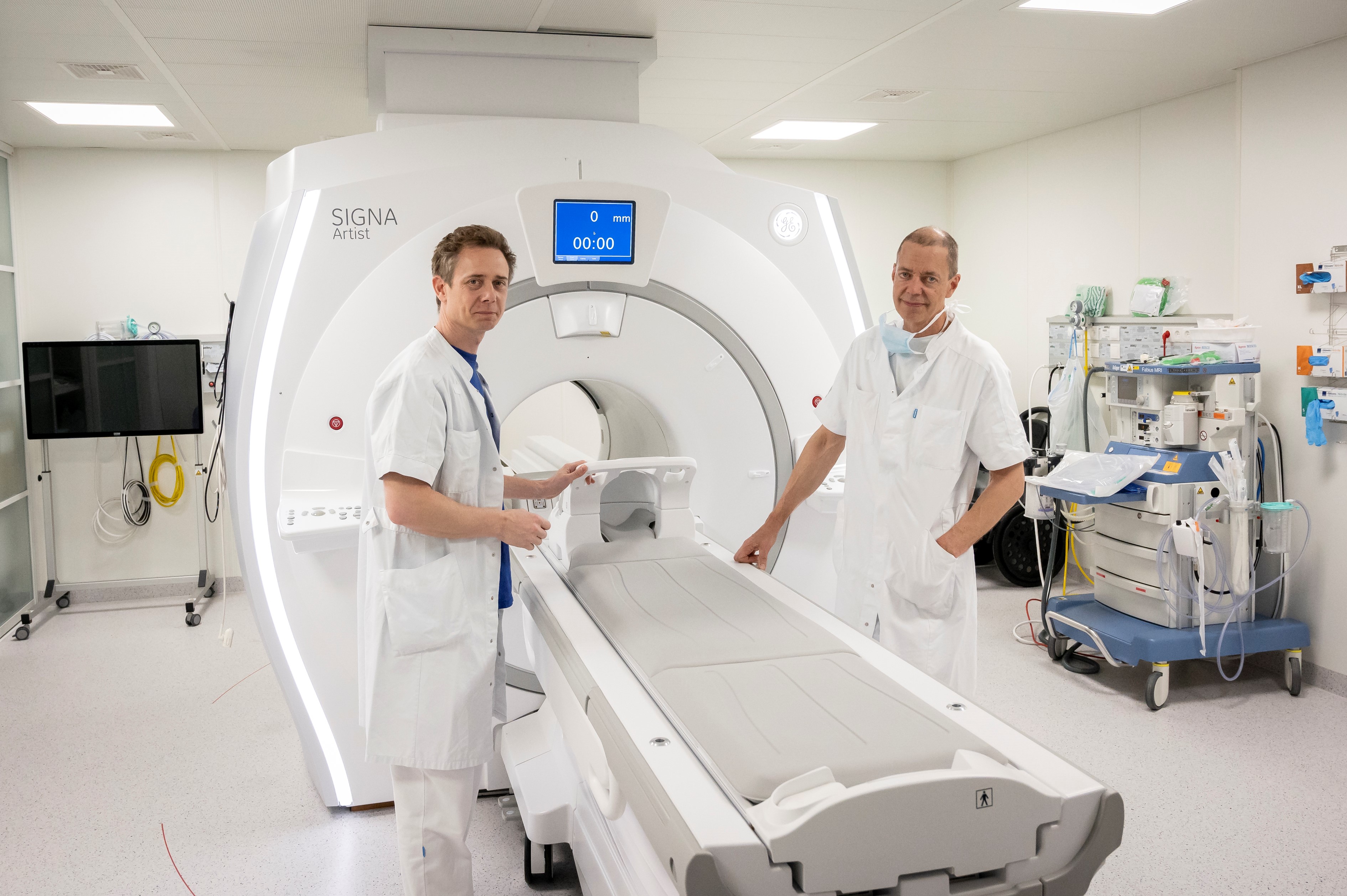
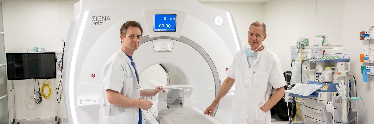
Neurosurgeons Rune Rasmussen and Bo Jespersen in front of their MRI scanner, the SIGNA™ Artist from GE Healthcare with the special Surgical Suite transport bed, on which a patient can be moved back and forth between the operating room and the scanner during brain surgery without moving the patient's head out of position.
MRI scans were traditionally used either for diagnosis and investigation or postoperatively to check whether the outcome of a surgical intervention was satisfactory. Now, MRI scanning can also be used during surgery as an integral part of the procedure. In the USA, MRI scanners have been used in direct connection with brain surgery for over a decade, and in recent years, the "intraoperative MRI" method has also started to gain ground in Europe.
Danish neurosurgeon and stereotactic neurosurgery specialist, Rune Rasmussen, is among the first in Europe to use MRI scanners during brain surgery, and we asked him to talk about his experiences so far.
What are the benefits of using an MRI scanner during brain surgery?
"Intraoperative MRI scanning is a prerequisite for a new surgical technique in which we ablate diseased tissue in the brain using an infrared laser. Intraoperative MRI enables us to monitor the temperature in the brain in real time, and thus control the treatment very precisely.
"This technique, called laser-induced interstitial thermal therapy or simply LITT, gives us the opportunity to perform surgical procedures on certain types of brain tumors and epilepsy with a greater degree of precision than before. In specific cases, we can even perform the procedure so gently that the patient can be discharged as early as the following day, which is quite a dramatic improvement from the patient's perspective.
"Generally speaking, this is a very good treatment option that we are pleased to be able to provide to selected patients."
”(...) LITT gives us the opportunity to perform surgical procedures on certain types of brain tumor and epilepsy with a greater degree of precision than before. In specific cases, we can even perform the procedure so gently that the patient can be discharged as early as the following day," says Rune Rasmussen, PhD, MD, Neurosurgeon.

Rune Rasmussen, PhD, MD, Neurosurgeon
You mentioned there are two ways to use an MRI scanner for neurosurgery: Moving the patient from the surgical table to the scanner and performing the procedure while the patient is in the scanner. What are the advantages?
"Yes, it is important to clarify that there are essentially two ways of using an MRI scanner in association with neurosurgical operations. The simplest is to move the patient from the surgical table and into the scanner once or several times during an operation. In the case of more sophisticated and complex operations, we perform the procedure while the patient is actually in the MRI scanner. It is called laser interstitial thermal therapy (LITT). That latter is the method I specialize in."
If we take the simpler operation first, moving a patient between an MRI scanner and the operating room in the middle of surgery immediately suggests a high risk. Why not wait until afterwards?
"It is not high risk. Our MRI scanner is located in a room between two operating rooms, each with direct access to the scanner. In the operating room, the patient is moved onto a specially designed transport bed that allows us to roll the patient into the scanner, carry out an examination, and then move the patient back onto the operating table, without moving the patient's head out of position. This means we can check whether, for example, we have removed all cancerous tissue from a tumor in the patient's brain or whether we need to remove more.
"The advantage of this is that we can be a little gentler. If we are unsure whether we should remove any tissue that is dangerously close to a vital area of the brain, we can now perform an extra scan to see if there is actually a need to remove this tissue as well."
"In the operating room, the patient is moved onto a specially designed transport bed that allows us to roll the patient into the scanner, carry out an examination, and then move the patient back onto the operating table, without moving the patient's head out of position. This means we can check whether, for example, we have removed all cancerous tissue from a tumor in the patient's brain or whether we need to remove more,” says Rune Rasmussen, PhD, MD, Neurosurgeon.
If we look at the more complex LITT technique, how is it even possible to operate while the patient is inside an MRI scanner?
"Specifically, the laser is controlled remotely via a screen, while I stand outside the room where the scanner and patient are located. As a prerequisite for me to be able to ablate a tumor using real-time monitoring with thermal images from the MRI scanner, the scanner must be carefully calibrated in advance with the equipment that is used to operate on the tumor. It takes a lot of preparation and is a more complex process overall."
And is it also faster compared to more classic open surgery?
"No, both simple and complex operations in which an MRI scanner is used during brain surgery are relatively resource intensive and involve a large team. The operation will involve one or possibly two surgeons, an anesthetist, two surgical nurses, two radiographers and support from the companies that supply the scanner and other equipment. And even if the actual procedure only takes an hour, as is the case with LITT, that's only the tip of the iceberg. The planning and preparation stages take a long time, which means the whole operation can easily take the best part of a day."
How important is it to have access to technical support from the MRI scanner supplier during this type of operation?
"I would say it is crucial. Access to expert support is needed before, during and after this type of surgery until we get to the point where we are so sure of the method and equipment ourselves that no longer need assistance to eliminate any risks or doubts regarding the technical setup, or the possibility of the equipment crashing in the middle of an operation. Fortunately, we have been very satisfied with the support we have received."
Can you describe some of the unique demands of LITT?
"We always want to be able to treat hard-to-reach lesions in the brain without causing unnecessary damage. The LITT method makes this possible, which is the very reason why it is so ingenious. In order to operate in the middle of the brain, it is necessary destroy some tissue on the way in — and the less tissue you have to destroy, the better.
"In addition to preparing and calibrating the equipment, it is this process of finding the least destructive route to the tumor and attacking it from the best angle that can be difficult and that takes time to plan. The laser ablation itself is not that difficult, but it is the part of the procedure where I am fully reliant on the support from the rest of the team, including from the radiographers operating the scanner."
Is there time savings associated with these MRI guided techniques and LITT?
"Not in the first instance, but this may well prove to be the case in the long run. Taking the 'simple' technique as an example, if a scan performed during an operation finds residual tumor tissue, this may mean that we can prevent the patient from having to undergo an additional operation, as we checked and ensured that all diseased tissue was removed during the procedure. This may well constitute a saving if you look at the balance sheet as a whole.”
"This may also apply to LITT treatment, although it is generally quite time consuming and resource intensive. However, it is also much gentler for the patient compared to open surgery, where we would go in hard, and where it also takes longer for the patient to recover.”
"When using LITT, we can simply drill a small 3-mm hole, which is enough to guide the laser catheter through. This explains why, in some cases, the patient can be discharged the day after such extensive brain surgery and therefore does not need to occupy a hospital bed for several days."
“If a scan performed during an operation finds residual tumor tissue, this may mean that we can prevent the patient from having to undergo an additional operation,” says Rune Rasmussen, PhD, MD, Neurosurgeon.
Is MRI-guided brain surgery a good treatment option for other patients, for example epilepsy patients?
"LITT is also highly relevant for epilepsy patients whose condition cannot be treated with medication and who may be severely disabled by repeated attacks. If we can locate the site in the brain where the epilepsy is originating, and if the relevant area is not too large, it can be ablated. In approximately fifty percent of cases, the treatment can completely cure epilepsy. Curative treatment is fantastic in every way and, of course, is completely life changing for the patient."
“In the context of epilepsy, it is believed that LITT may replace a lot of open surgery in the long run,” says Rune Rasmussen, PhD, MD, Neurosurgeon.
You mentioned a number of benefits, but are there also limitations?
"Yes, there are, in spite of everything. If a tumor is larger than a table tennis ball, the heat from a laser catheter cannot spread far enough into the tissue, so we have to resort to the knife again. It is important to make it clear that, as it stands, the LITT technique is not about to replace standard tumor surgery. But it is a fantastic supplement — a new tool in the surgical toolbox that can be offered to a specific group of patients with brain tumors. On the other hand, in the context of epilepsy, it is believed that LITT may come to replace a lot of open surgery in the long run."