A new approach for CTO using CCTA
Interview with Paul Handwerk (PH), radiographer at the Saint-Georges clinic, since 2020 and Doctor Laurent Drogoul (LD), interventional cardiologist.
"We perform around 3500 procedures per year including 1500 dilations and around 80 CTOs.”
Can you describe the complementarity between radiographer and cardiologist in your daily practice?
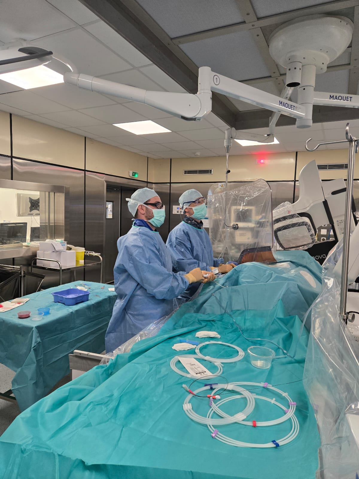
Paul Handwerk: As radiographer, we are fortunate to be able to assist the doctors and to be part of the exam with them. We are responsible for setting up the patient prior to the exam and during the procedure we are the ones who handle the table and the C-arm. By being close to the procedure, we can also help in the preparation of the devices. For complex procedures we are helped by a nurse to provide the required equipment.
Laurent Drogoul: The specificity in our clinic is that the radiographer manipulates the angiography system and scrubs in with us for angioplasties. There is great complementarity in the cardiologist/radiographer pairing since with practice, we will be able to discuss the device and the strategy to adopt for complex procedures, in particular CTO. Furthermore, as the radiographer oversees the system and follows the entire procedure, they can anticipate our needs (material, angulation, etc.) which allows us to gain in efficiency since we remain completely focused on the technicality of the procedure.
What are your problems in your Cath Lab during complex procedures? And how do you manage it?
Paul Handwerk: Our main problem is to clearly identify the diseased areas, the type of plaque and to anticipate the complexity of the exam before starting it to prepare for all eventualities. This makes it possible to adapt the treatment and the type of device that will be used (stent or not, active balloon, etc.). In the case of CTO for example, we are limited by the cost of certain technologies (IVUS, OCT, etc.) hence the interest in working on the Coronary CT Angiography (CCTA) of the patient to obtain the maximum information on the pathology. The advantage in our clinic is that cardiologists perform CCTA and can therefore provide indications for coronary angiography.
Laurent Drogoul: What we need most is to go beyond the angiographic image. We need to see the calcifications and quantify them. Looking at the internal anatomy of the vessel, are the calcifications circumferential? Its length, its diameter. For example, in CTO, the anatomy is ambiguous, we do not necessarily know where we are and angiography does not allow us to answer the question. The informations provided by tools such as IVUS and OCT are interesting but those require too much contrast and/or are still too expensive. The CCTA is therefore an interesting imaging test to answer some of our questions.
Can you describe the patient journey for CTO procedures in your facility?
Laurent Drogoul: There are two main paths. The first is in our center either via a diagnostic coronary angiography for symptoms or following a CCTA. The second is patients who are referred to us from other centers because they are complex cases and we are a reference center in the region. Today, due to time and organization, there are still many patients who arrive in the cath lab without a CCTA, but I think that this will change in the years to come since it becomes the diagnostic exam of reference.
You tried an innovative technique based on the use of CT to help you in the cathlab, could you describe it?
Paul Handwerk: The objective for us is to obtain a co-registration between the cath lab and the CT-Scan. For this, we have on our AW station all the software which allows the automatic segmentation of CCTA (image 1). In addition, this station is directly connected to our IGS room. We are therefore able to use the 3D reconstruction as a mapping of the patient’s coronaries and observe the trajectory of the CTO to anticipate the guide wire’s path. The 3D modeling of the scanner is displayed directly on our large display monitor in the room, respecting the scale of the anatomy (image 2). The doctor is able to take measurements and make annotations (image 3). The other advantage is that when the C-arm moves, the 3D model follows the orientation of the imaging system. We have the same incidence between the angiographic image and the CCTA. This allows you to choose the optimal angulation and visualize the trajectory of the CTO from the CCTA.
Laurent Drogoul: When we do a double injection for a CTO, we cannot visualize the occluded area. We can only imagine it mentally. The CCTA provides us with this information on the appearance of the lesion and its possible tortuosity. This valuable information allows us to direct our guide when we unclog the coronary artery and allows us to be sure that we are in the right direction.
| Image 1 | Image 2 | Image 3 |
Do you think such a technique could become part of your routine for these procedures?
Paul Handwerk: Everything will depend on the availability of the CCTA. All patients who have had a screening scan, I think it is important to use it because there are studies1, 2 which show that it has a beneficial effect for the patient. Then there are CTO classifications. For those classified as simple, the question arises about the benefit/risk of irradiating the patient knowing that we will have a high clearance and success rate in these procedures. For complex CTOs, it is up to the doctor to judge but I think it is a real benefit to have a CCTA to help during the procedure.
Laurent Drogoul: I think that the CCTA is becoming an essential imaging test. It allows us to measure the length of the CTO, the diameters of the artery, anticipate the pitfalls... Where angiography will never be able to fully answer these questions. We are therefore counting on manufacturers and GE HealthCare to continue to innovate in cross modal imaging to always better see the coronary anatomy.
1 Accuracy of J-CTO Score Derived From Computed Tomography Versus Angiography to Predict Successful Percutaneous Coronary Intervention
2 https://pubmed.ncbi.nlm.nih.gov/33550529/
M Paul Handwerk and Dr Laurent Drogoul have not been paid by GE HealthCare for this testimonial. The statements by M Paul Handwerk and Dr Laurent Drogoul described here are based on their own opinions and results that were achieved in their unique setting. Since there is no “typical” hospital and many variables exist, i.e. ambulatory surgery center size, case mix, etc. There can be no guarantee that other customers will achieve the same results.
Case Report CCTA
Patient
71-year-old patient with primary implantation of a dual-chamber pacemaker following BAV III. He also performed a CCTA with a calcium score of 1555. He also has risk factors: tobacco, 50P/A hypertension and dyslipidemia. We therefore perform a coronary angiography where we find a CTO on the LAD (image 1) and significant stenosis of the distal circumflex artery (Cx) and moderate plaques in the right coronary artery (RCA). Following this exam, an ultrasound and a cardiac MRI showed viability. It is therefore decided to dilate the CTO on the AIV and the Cx.
Technique
We went for a double radial approach CTO with 7f anterograde and 5f retrograde because the LAD is given by the RCA (image 2). Considering the CCTA (length of the lesion, complexity of the anatomy) we chose an anterograde approach (image 3). We used a Gaia microcath 1 then 2 to pass the lesion. Considering the angiographic images and the CCTA, we pre-dilatated with a balloon to be sure to open the heavily calcified areas. We placed a long stent with a diameter of 2.5mm (consistent with the measurements taken on the CCTA) (image 4). Then we placed a 2nd stent with a diameter of 3.5mm (still consistent with the measurements taken with the CCTA). We then treated the bifurcation with the diagonal by pot/kiss/ pot. Finally, we used StentViz (image 5) to ensure the proper deployment of the stent, particularly in the calcified areas where we observed a good result.
Benefits of CCTA
- Analysis of the anatomy and tortuosity of the CTO, length and diameter measurements, annotations, etc.
- Visualization of the anatomy directly on the large display to choose the best working angle.
- Help in the choice of device and its orientation in a complicated anatomy.
Procedure information: 160mL of iodine, 152,2 Gy.cm² and 29 min of fluoroscopy.
Image 1
Image 2
Image 3
Image 4
Image 5
© 2024 GE HealthCare. GE is a trademark of General Electric Company used under trademark license. All rights reserved. The statements by GE HealthCare customers described here are based on their own opinions and on results that were achieved in the customer’s unique setting. Since there is no “typical” hospital and many variables exist, i.e. hospital size, case mix, etc., there can be no guarantee that other customers will achieve the same results. JB07969XE