Dr. John Pak and Dr. Dan Christensson, consultants in spine surgery, provide their perspectives on OEC 3D, illustrated through their experience during a case of spine arthrodesis.
The hospital of Halmstad from the county of Halland is located on the southern west coast of Sweden that offers beautiful nature comprising of beaches, forest and lakes. Serving the county to population of 300,000, Halland Hospital Halmstad offers a full range of specialized care to the population. Thus the orthopedic clinic of Halland Hospital Halmstad has developed a unit dedicated to spine surgery which launched the process of evaluating mobile C-arms for 2D and 3D intraoperative imaging.
The main motivation for the acquisition of a 2D/3D mobile C-arm is to obtain, in addition to traditional 2D images, comprehensive 3D volumetric images in the operating room for intraoperative clinical decision making. The team also wanted to assess the possibility to streamline the patient’s pathway by eliminating the postoperative CT scan confirmation, based on the 3D imaging control performed in the operating room at the end of the intervention.
L4-S1 fusion by posterior approach with 2D and 3D imaging
Courtesy of Dr. Nils Petter Fossland and Dr. Mats Säll, St Olavs University Hospital, Trondheim, Norway
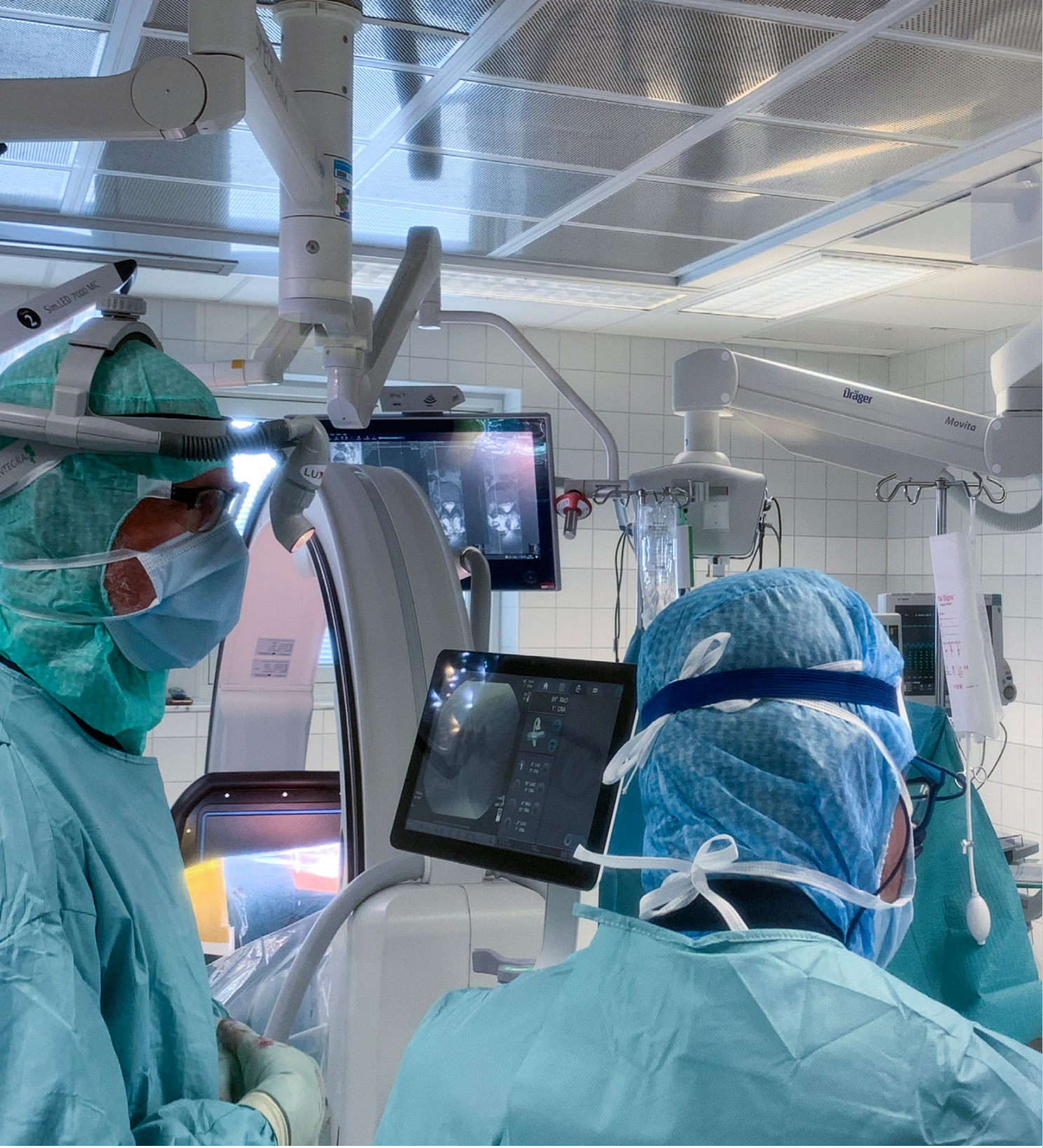
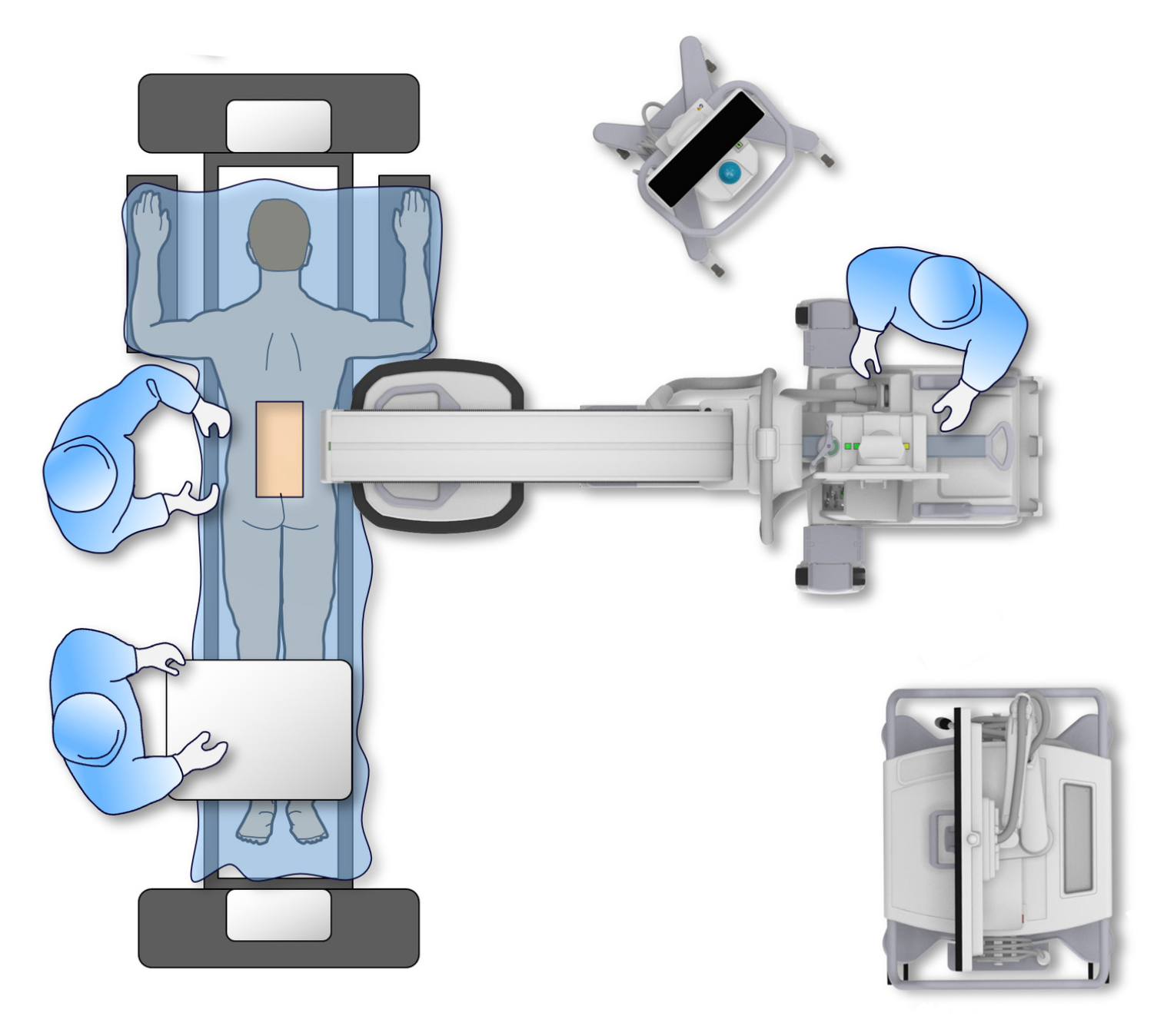
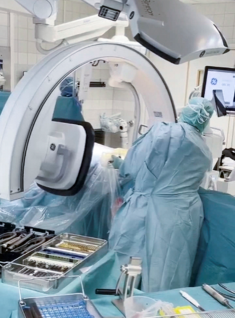
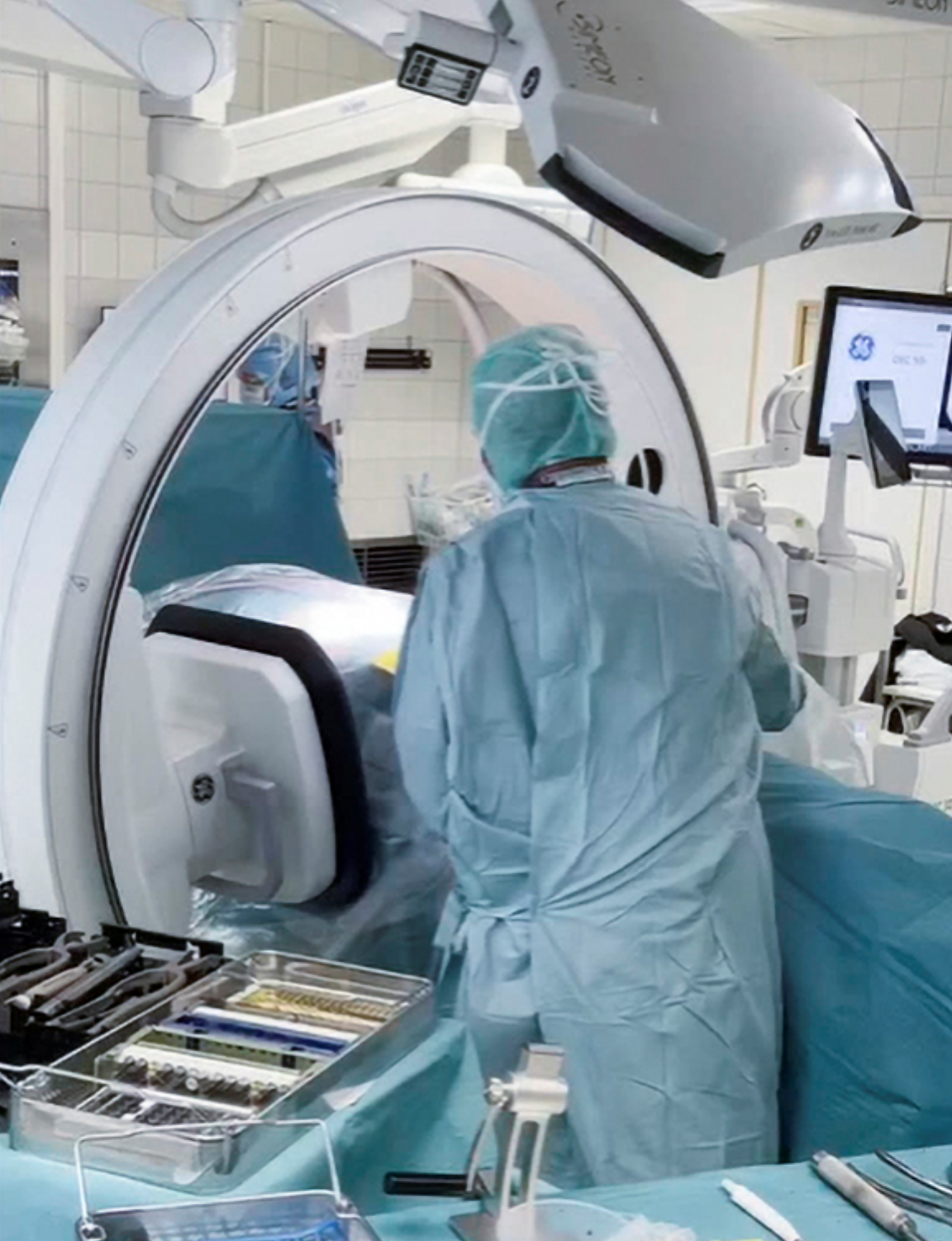
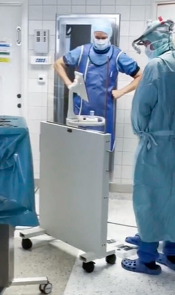
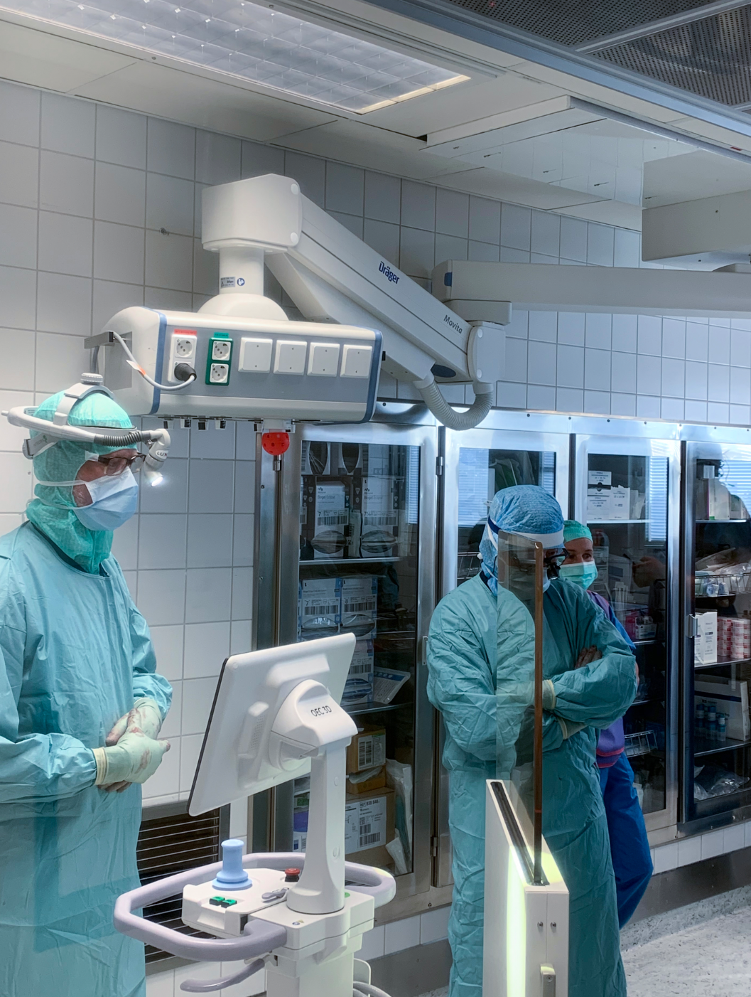
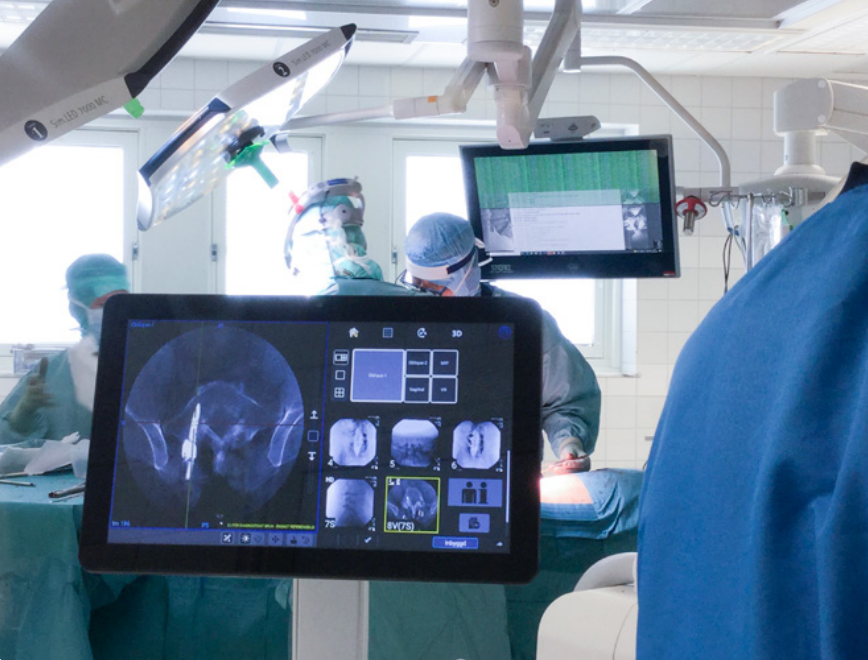
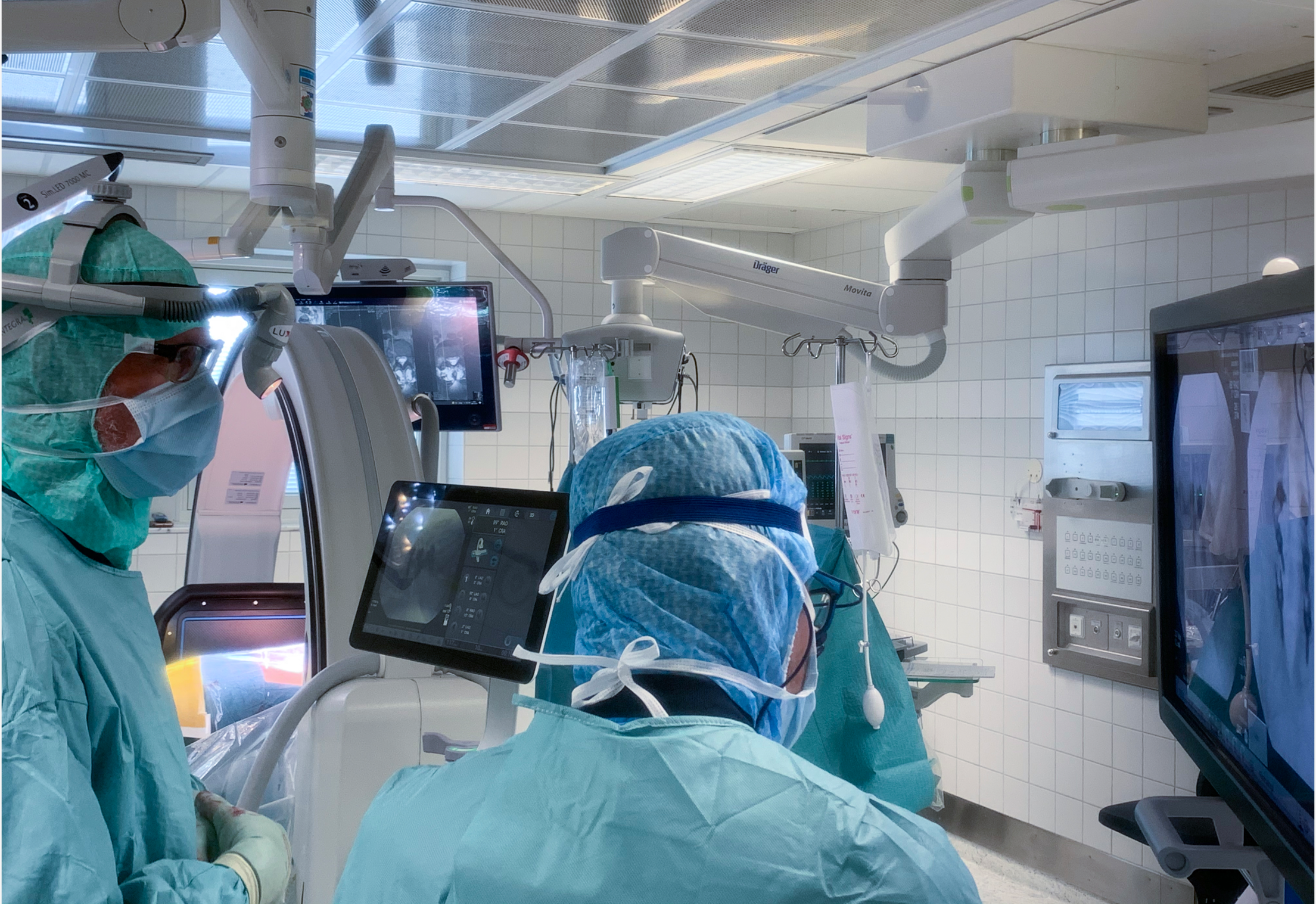
Patient and C-arm Set up
Patient is positioned in prone. The C-arm is placed perpendicularly to the patient table.
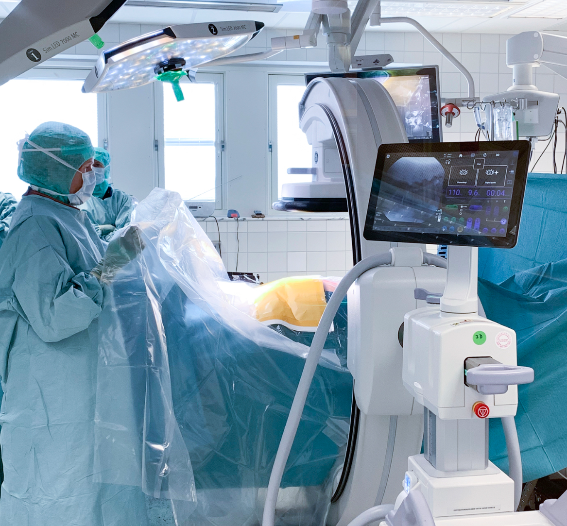
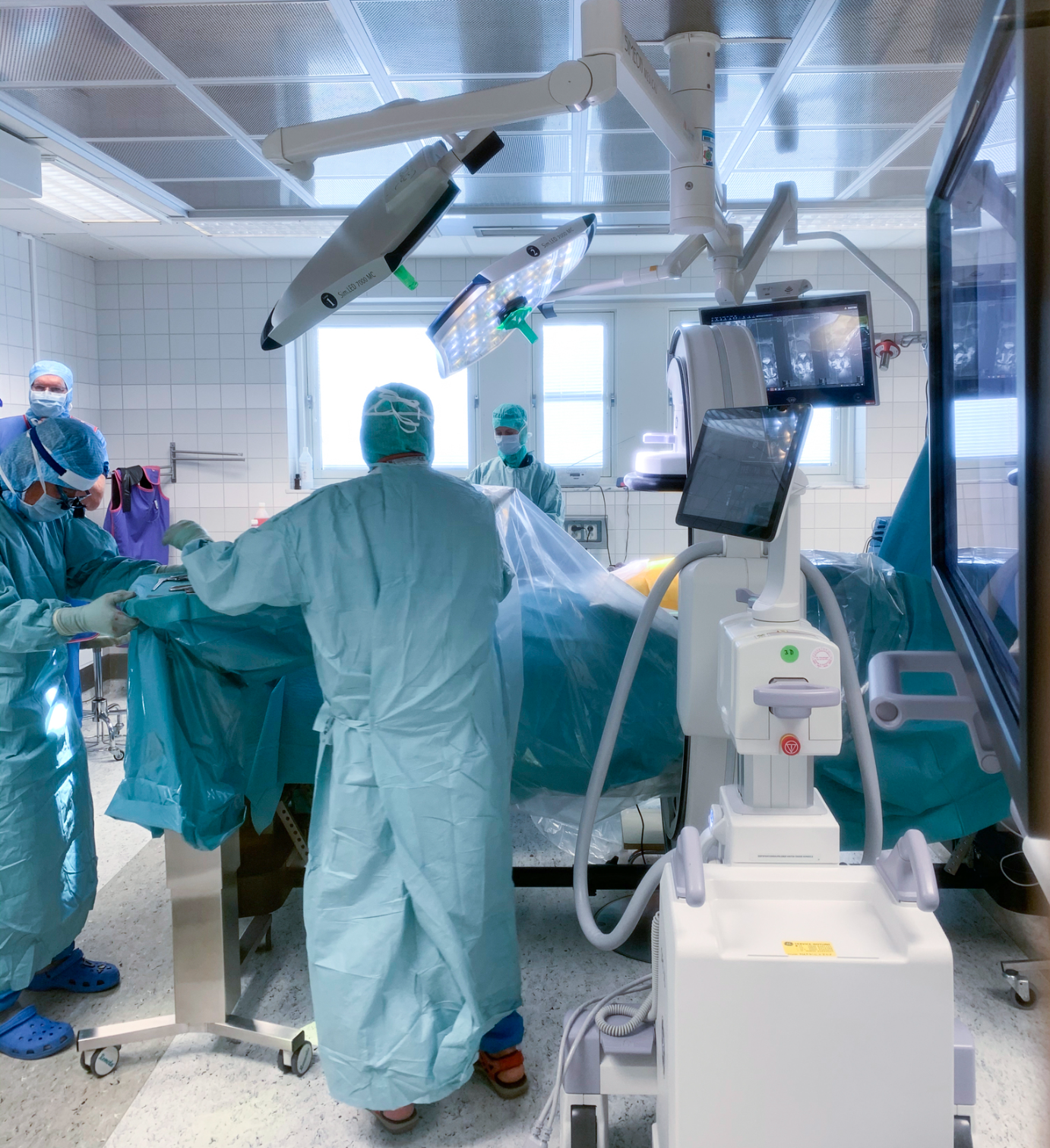
Operating room set-up for spine 3D images acquisition
3D Imaging Every Day
The OEC 3D is a true 3D/2D C-arm with the flexibility to acquire both precise 3D and 2D images without switching systems. Transitioning quickly and easily provides greater efficiency and versatility for a wide range of clinical applications from spine and orthopedics.
Precise Images
The OEC 3D enables allows to see more levels during a spinal fusion, or more of the pelvis or femur during an orthopedic procedure. With a 19 cm x 19 cm x 19 cm volume, OEC 3D captures a 67% greater volume than other 3D C-arms*.
The 3D acquisition designed for capturing as much data as possible is critical in creating highly detailed images. The OEC 3D delivers a true 200° isocentric orbital sweep, 35° more than other 3D C-arms*, for comprehensive 3D volumetric images.
The OEC 3D leverages GE Healthcare’s proven AW image fabric technology to provide a premium 3D imaging experience. Analyze images with the Volume Viewer suite of 3D imaging tools including Multi-Oblique Mode, viewing all five image perspectives (Axial, Sagittal, Coronal, Volume Rendering, Maximum Intensity Projection) through all 512 slices, window leveling, zoom, and more.
*Compared to other 3D C-arm published specifications
2D intraoperative imaging
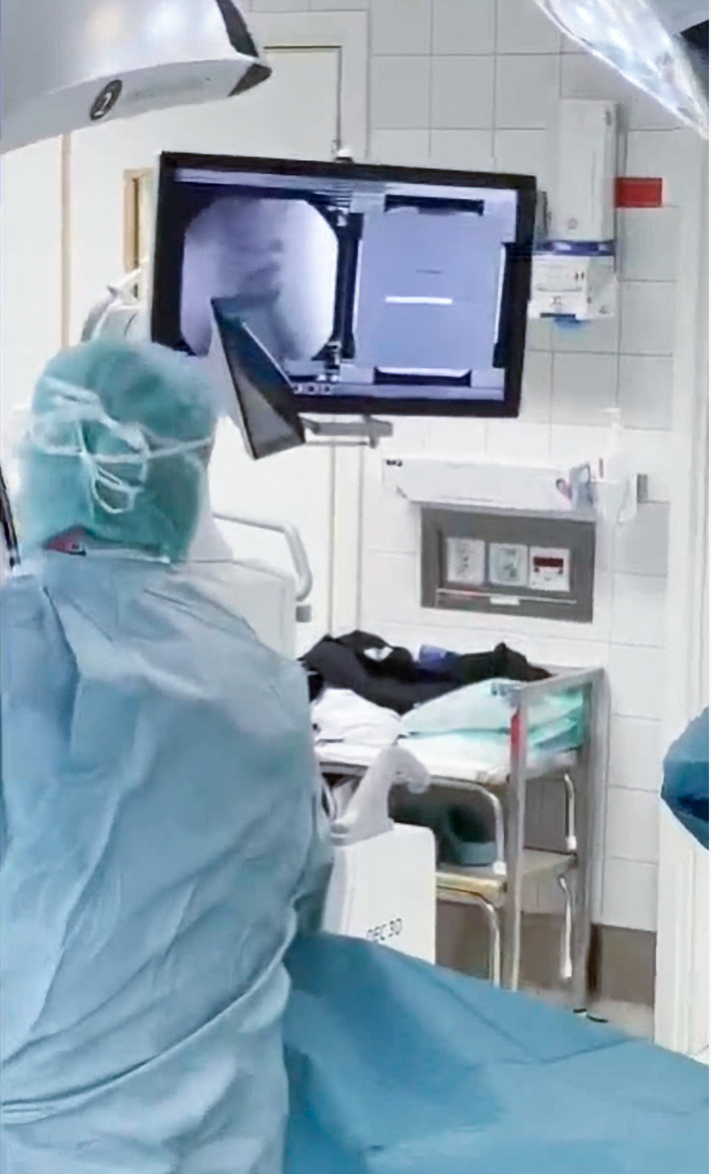
Anatomical imaging profile is set to Spine. Fluoroscopy mode is set to standard continuous. A first fluoroscopic image is taken to confirm the vertebral level to treat. Further 2D imaging centering is done using the Live View camera of C-arm to limit the number of image acquisition and manage dose radiation level.
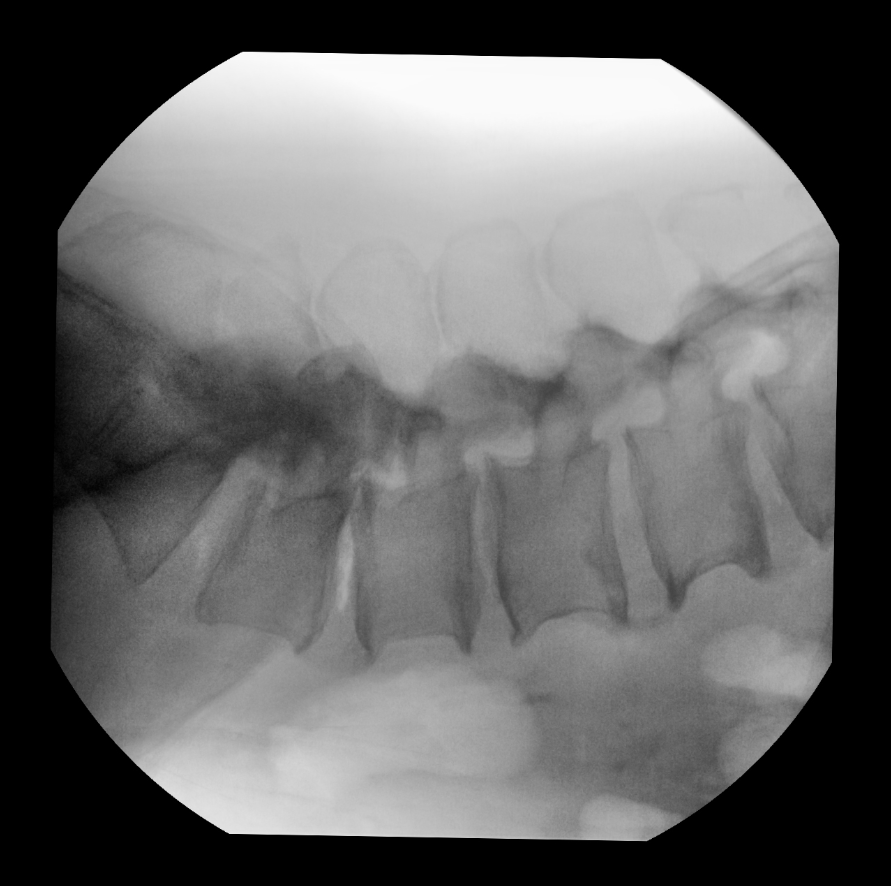
Lateral view – definition of incision level on L4
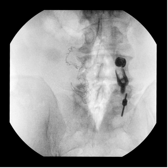
Antero Posterior view
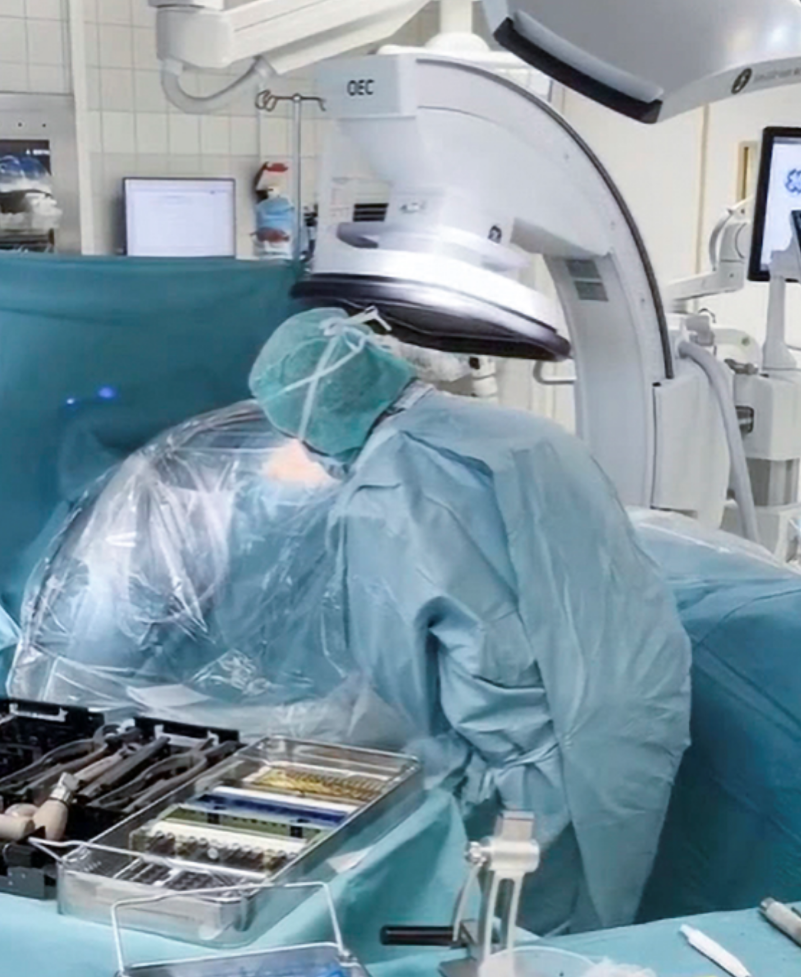
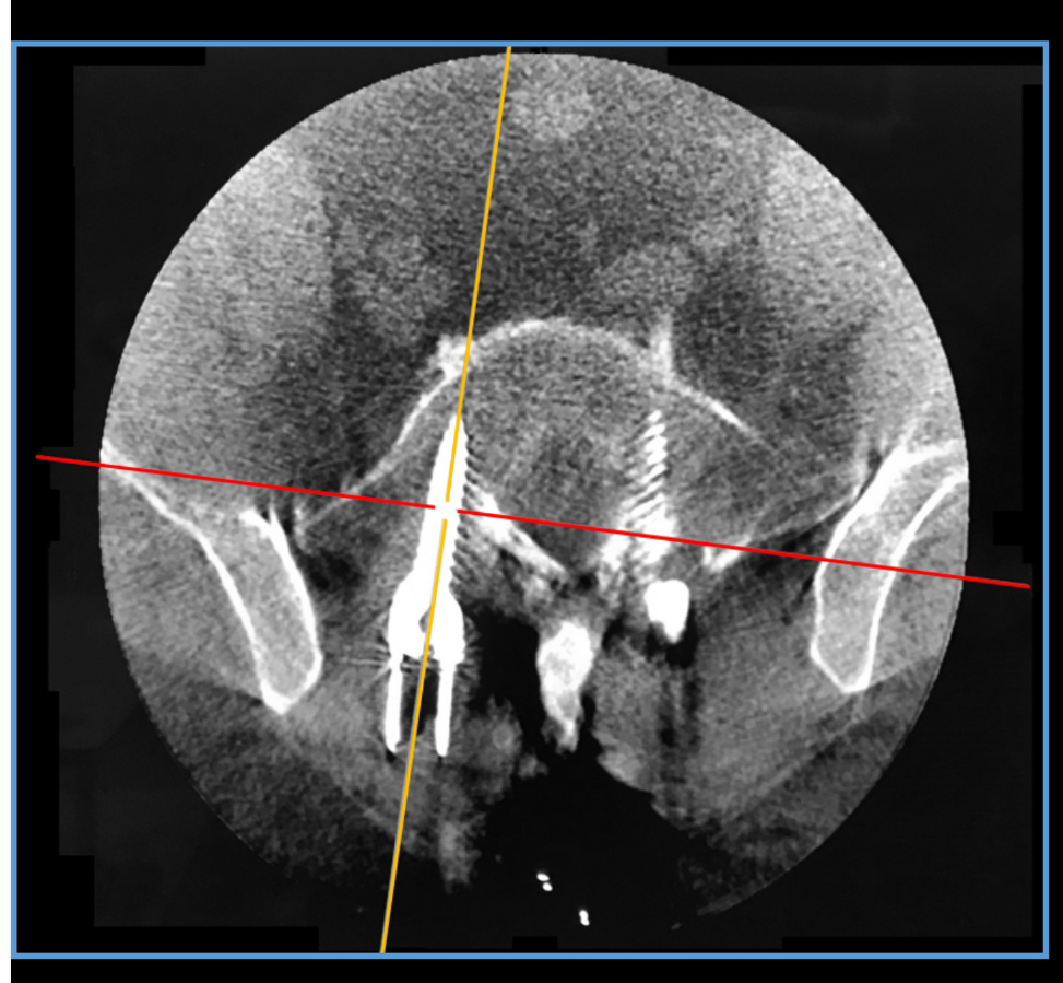
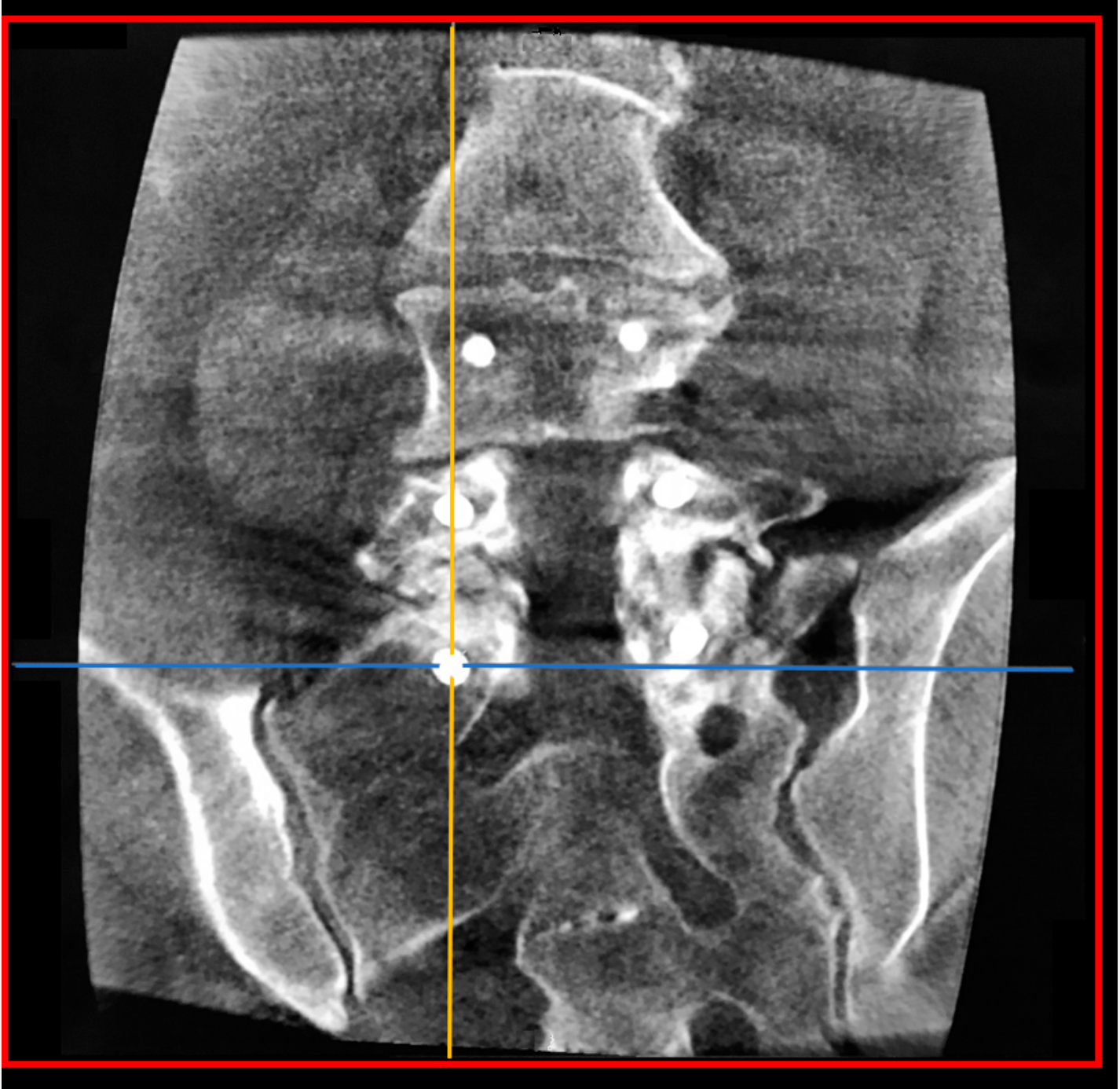
Once all the pedicle screws are placed, surgeons proceed to the 3D acquisition on L4-S1 to control the overall surgery on volumetric images.
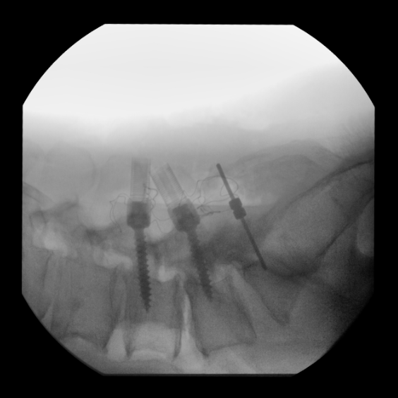
Lateral View
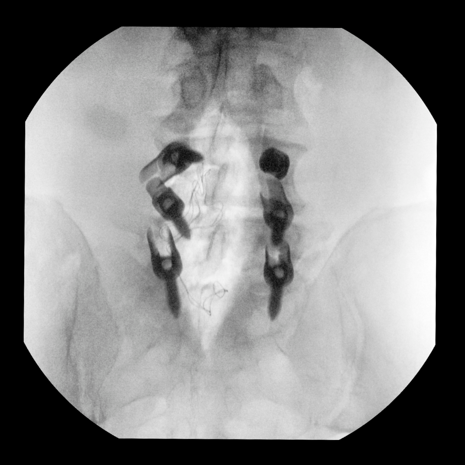
Antero Posterior View
3D imaging final control with Setup Assistant
The Setup Assistant guides the user through 4 steps to acquire the 3D scan. After setting the 3D imaging plan and centering the anatomy in the 3D volume, the Setup Assistant allows the user to check that the C-arm rotates freely around the patient.
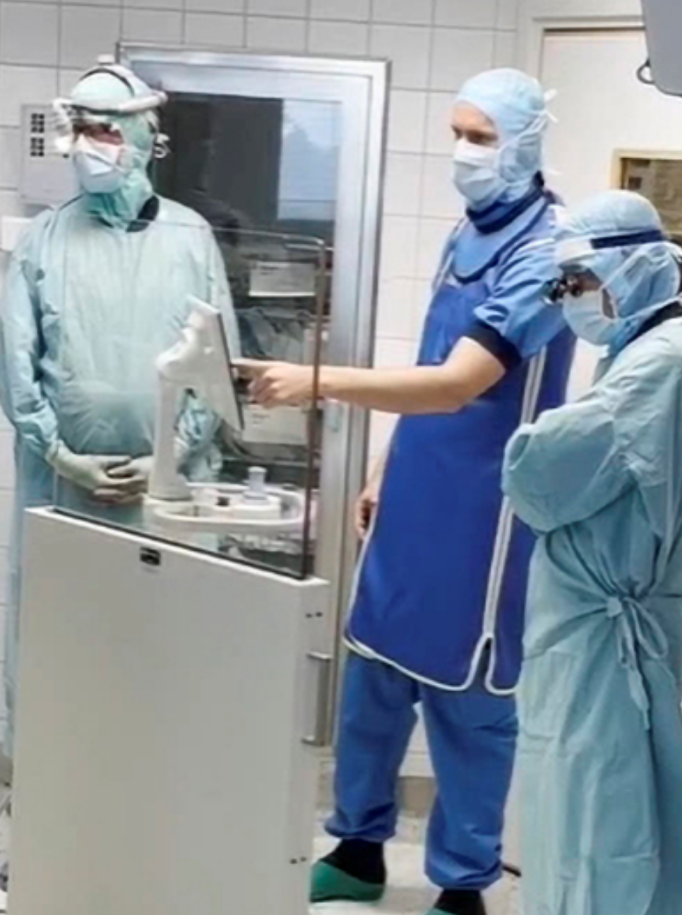
“I could make the operation with quick and easy 3D imaging”
Dr. John Pak
3D scan acquisition from wireless pedal footswitch
At the end of the 3D scan, the data set is reconstructed and presented on displays for viewing in the Volume Viewer for precise analysis of 3D perspectives.
“I feel more confident to precisely control the position of the screws in the vertebral pedicles before closing the patient. It helps me to assess the quality of my surgical gesture to avoid revision surgery.”
Dr. Dan Christensson
The Volume Viewer presents high resolution 3D images, from the 19 cm x 19 cm x 19 cm volume, in five perspectives. The volume visualization is based on AW image fabric technology to provide a premium 3D imaging experience. User an analyze images with the Volume Viewer suite of 3D imaging tools including Multi-Oblique Mode, scrolling through all slices, window leveling, zoom, and more.
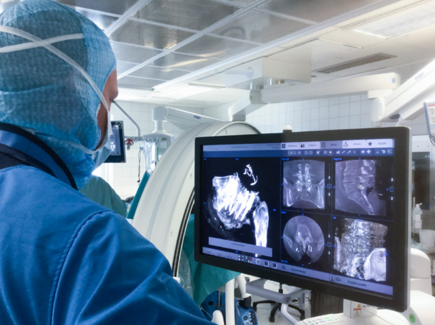
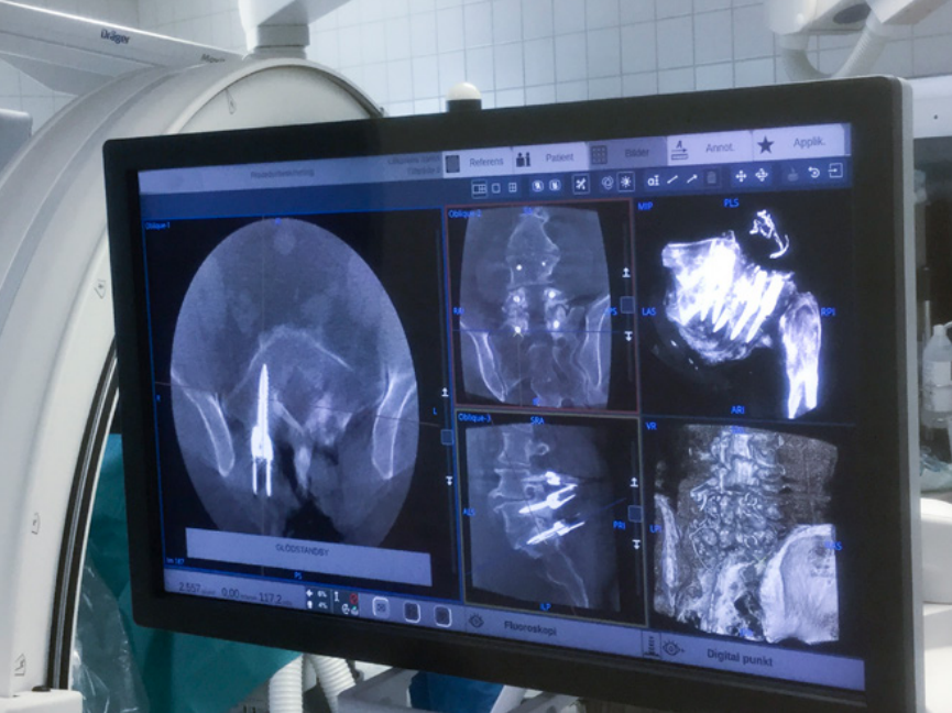
Each view can be placed on the left side for larger format visualization.
“With OEC 3D you can see all the vertebras at the same time, it’s fantastic! It has a wider field of view and more visualization power.”
Dr. Dan Christensson
“The Image Quality of 2D and 3D images is very good even on obese patients. It is important because people are getting more and more obese and we have to deal with more soft tissues, which bleed more and it’s becoming more complex to see.”
Dr. John Pak
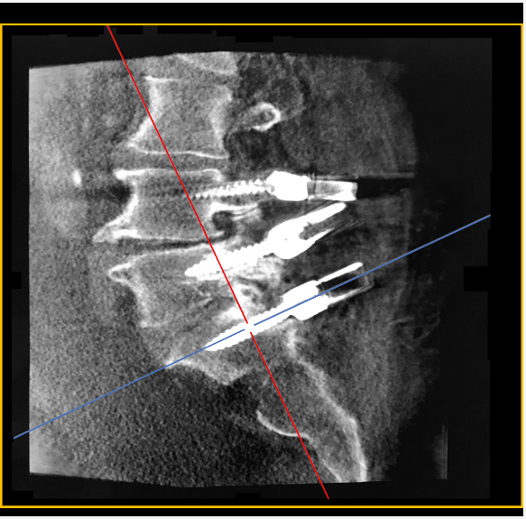
Oblique views of L5 left pedicle screw
Conclusion
The clinical benefit of 2D and 3D imaging with OEC 3D C-arm delivering precise images in a 19 cm x 19 cm x 19 cm volume led the team to select OEC 3D C-arm for their daily practice.
“OEC 3D is full of options and has capacity above our needs.”
Dr. John Pak
DOWNLOAD THE FULL 3D OEC MAGAZINE #5
gehealthcare.co.uk/-/jssmedia/gehc/uk/files/products/surgical-imaging/oec-magazine5high-res.pdf?rev=-1
The statements by GE’s customers described here are based on their own opinions and on results that were achieved in the customer’s unique setting. Since there is no “typical” hospital and many variables exist, i.e. hospital size, case mix, etc.. there can be no guarantee that other customers will achieve the same results. JB05583XE