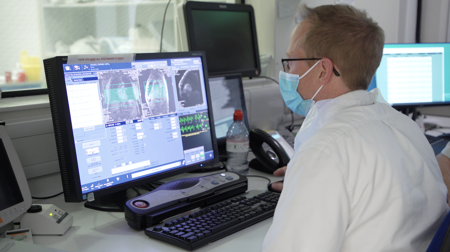
Cardiovascular disease causes a quarter of all deaths in the UK and is the single largest condition where the NHS could save lives over the next decade[1]. Historically, cardiologists have relied predominantly on subjective methods of valvular heart disease assessment, typically echocardiography, which is limited to imaging one-directional flow. A more recent development is 4D flow cardiac MR, a complementary technique offering a multidirectional, real-time view of blood flow that allows cardiologists to pinpoint heart valve leaks and make more informed decisions about a patient’s treatment.
Specialist cardiology is a key area for the Norfolk and Norwich University Hospital (NNUH), which is currently undergoing an £8m project to replace ageing CT and MRI scanners and establish state-of-the-art diagnostic methods, including the introduction of 4D flow cardiac imaging. Dr Pankaj Garg, a Consultant Cardiologist and Associate Professor at the University of East Anglia, explained: “NNUH is a teaching trust hospital with a strong focus on research, always seeking to translate novel techniques into clinical practice, to improve services and health outcomes, and the patient experience. The experience I gained in cardiac MR and 4D flow research at the Universities of Leeds and Sheffield is helping us to introduce this cutting-edge imaging technique into clinical practice at NNUH. We’re the first hospital in the UK to offer 4D flow cardiac MR.”
Complementary technologies help show the bigger picture
Historically, cardiologists have relied on echocardiography for valvular heart disease assessment. Initially this would be a transthoracic echocardiogram, followed, if necessary, by a transoesophageal echocardiogram (TOE) to define the morphology of the valve and assess the leak. However, TOE is an invasive procedure requiring sedation – not ideal, especially during the COVID-19 pandemic – and cannot be performed on all patients. This meant that, until now, a significant proportion of patients did not have the benefit of additional investigations to further explore the severity of their condition prior to embarking on surgery. 4D flow cardiac MR helps overcome this issue.
Richard Greenwood, MRI Deputy Lead and MRI Research Lead Radiographer, commented “4D flow cardiac MR gives us huge amounts of additional information that we wouldn't otherwise be able to take advantage of, which obviously improves the pathway for the patient. It's a non-invasive procedure and fits in nicely with our clinical cardiac MRI protocol.”
Dr Garg added: “Complementing echocardiography with standard cardiac MR goes so far, offering more precise quantification. For example, mitral regurgitation is generally evaluated by quantifying the aortic net forward flow through the aortic valve using phase contrast acquisition methods, along with cine acquisition of the left ventricular stroke volume; the rate of regurgitation is determined by subtracting one from the other. However, this involves two different acquisitions, and there is a spatial misalignment issue, leading to a degree of imprecision. This is where 4D flow cardiac MR takes the level of detailed information one step further, providing both temporal and spatial coverage.”
Enhanced information
Introducing 4D flow cardiac MR into clinical practice provides an additional level of confidence in reporting the severity of mitral valve leakage. Dr Garg described the benefits: “The ability to acquire directional information across four dimensions makes 4D flow cardiac MR a much more accurate and reproducible tool than those used previously, complementing other technologies to enable a more detailed examination of the heart for improved diagnostic accuracy. Vast amounts of data – between 18 and 20,000 images – can be acquired in just a few minutes, with no additional patient interventions required. This enhances the quantification of valvular disease, to guide patient management moving forward.”
More informed decision-making
Accurate assessment of the severity of valvular incompetence is essential to determine the best course of patient treatment and effectively manage the condition. NNUH is already seeing clear benefits from using 4D flow cardiac MR as an adjunct to standard cardiac MRI and cardiac echo flow measurements. Dr Garg continued: “4D flow cardiac MR enables a more in-depth analysis of the patient’s condition, giving clinicians extra confidence in their diagnoses. In some cases, it has even allowed me to eliminate mitral valve incompetency as the cause of the patient’s problem, identifying a different clinical issue – for example cardiac amyloidosis or cardiac sarcoidosis – that did not require surgery. These instances have been hugely significant; without the results of the 4D flow cardiac MR, these patients would almost certainly have gone on to have an interventional procedure that would have been completely unnecessary. This has tremendous repercussions for the patient, as well as on resources.”
Every minute counts
NNUH uses a 3 Tesla MRI scanner from GE Healthcare and new 4D flow cardiac MR software to image the heart valves precisely and cost-effectively as part of a standard cardiac MR, without prolonging the examination time. Dr Garg explained: “During a routine cardiac MR there is a lag time between the administration of gadolinium contrast agent and performing the late enhancement scan. 4D flow cardiac MR using a HyperKAT sequence – a GE Healthcare patented acceleration technique – fits easily into this period, making full use of the available MR time without significantly affecting the overall length of the patient’s appointment.”
Providing the best patient experience
Patients are understandably concerned when a potential heart problem is identified, and assessing the severity of any condition non-invasively, minimising patient discomfort and worry, is clearly preferable. “The patient impact of 4D flow cardiac MR could be quite phenomenal,” remarked Dr Garg. “Patients realise that they are benefitting from cutting-edge technology to answer critical questions for their healthcare, and they are excited to be at the forefront of modern medicine. 4D flow cardiac MR is a far more patient-friendly alternative to other techniques used to assess valvular incompetency. This non-invasive, free breathing, non-respiratory navigated procedure fits into a standard cardiac MR workflow without extending the time spent on the scanner – which is particularly important for patients who are claustrophobic – enhancing the overall patient experience.”
“The patient experience will be further improved in the near future, as we are shortly due to upgrade to SIGNA™ Premier AIR™, which offers even faster acquisition times to increase throughput, and lighter coils that will be more comfortable for the patient,” added Rayhaan Rahaman, Consultant Radiologist and Chief of Imaging.
Blazing a trail
The translation of 4D flow cardiac MR from research into clinical practice has positioned NNUH as a leading provider of cardiac imaging in the UK, and the hospital is proud to be the first clinical site in the country to implement this advanced diagnostic tool, revolutionising the patient pathway for people with valvular heart disease.
[1] www.nnuh.nhs.uk (n.d.). We’re first to use 4D flow MRI scans to help patients with heart disease. [online] Available at: https://www.nnuh.nhs.uk/press-release/were-first-to-use-4d-flow-mri-scans-to-help-patients-with-heart-disease/

