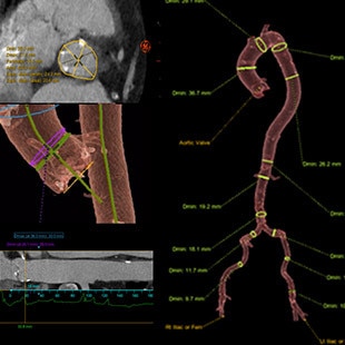
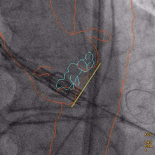
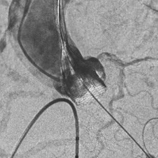
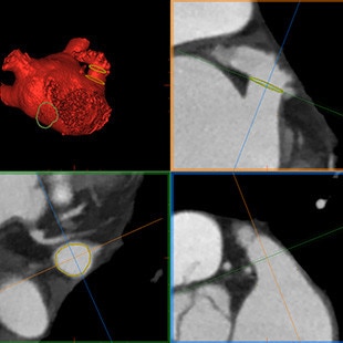
TAVI

Transcatheter Aortic Valve Implantation

Plan

Choosing the right size of the valve is key because inadequate sizing has been identified as a predictor of paravalvular leaks1

Valve ASSIST 2(4)

0-click aorta segmentation and valve definition makes accurate valve sizing accessible to every one


GUIDE

Precise positioning of the aortic valve is key to avoid leaks & regurgitations2

Valve ASSIST 2(4)

Direct import of planning lines in the cathlab, overlaid with X-Ray to simplify navigation

Calcification enhancement improves the visualization of moving contrasted structures


ASSESS

Assessment of device deployment and regurgitation is essential, because moderate / severe aortic regurgitation is associated with a 72% mortality risk3 and malpositioning of the valve being identified as a predictor of paravalvular leaks1

Valve ASSIST 2(4)

Assess potential regurgitation with exceptional contrast visualization


LAAC

Left Atrium Appendage Closure

Plan

Understanding the anatomy of the left atrium and shape of appendage is important

Valve ASSIST 2(4)

Segment the 3D CT images of the left atrium to precisely plan the intervention

Import planning lines in the cathlab, overlay with X-Ray to simplify navigation

Improved flexiblity
Ability to import DICOM data from other planning software tools


GUIDE

Precisely positioning the device can be challenging and has an impact on complete sealing

Valve ASSIST 2(4)

Better visualize the left atrium & appendage geometry to guide the procedure


In LAAC procedures7:
-78% contrast media volume
-28% procedure time
-25% fluoroscopy time


INTERACT View X(5)

Enables display of the live X- Ray image on the U/S system

Precisely guide device delivery inside the 4D volume in real-time


ASSESS

Device deployment assesment & assessment of regurgitation

Vivid™ Imaging(6)

Confirm with echocardiography the device location and evaluate compression before releasing the device


TMVR

Mitral Valve Repair and replacement

Plan

Planning phase is key to the intervention and reducing LVOT obstruction

Cvi42 mitral(8)

Easy, efficient anatomical analysis of the mitral valve for pre-procedural planning transcatheter mitral valve interventions


4D Auto MVQ on Vivid™ Imaging(5,9)

Rapid semi-automated detection of the mitral valve morphology, including the annulus and the leaflets

Stores of complete setup and results together with the exact screen view for quality control


GUIDE

Precisely positioning the device can be challenging and has an impact on regurgitation

Vivid™ E95(6)

Provides exceptional volume rates in single beat imaging helping to safely guide device deployment

FlexiSlice in-live allows the precise positioning of every single screw to be guided during the procedure


ASSESS

Device deployment assesment & assessment of regurgitation

Vivid™ Imaging(6)

Assess potential regurgitation with excellent temporal and spatial resolution


TTVR

Transcatheter Tricuspid Valve Repair and replacement

Plan

Planning phase is key to the intervention

Valve ASSIST 2(4)

Segment the 3D CT images of the left atrium to precisely plan the intervention, including:
• Analysis of the morphology
• Localization of ideal landing zone
• Anchorage point localization


GUIDE

Precisely positioning the device can be challenging and has an impact on regurgitation

Vivid™ E95(6)

Provides exceptional volume rates in single beat imaging helping to safely guide device deployment

FlexiSlice in-live allows for guiding the precise positioning of every single screw during the procedure deployment


ASSESS

Device deployement assesment & assessment of regurgitation

Vivid™ Imaging(6)

Rapid evaluation of procedural success and help for diagnosis of complications


See also the main challenges in Coronary Angiography

READ MORE

Heart Care

  1. Paravalvular Leak After Transcatheter Aortic Valve Replacement. The New Achilles’ Heel? A Comprehensive Review of the Literature - https://www.ncbi.nlm.nih.gov/pubmed/23375925
  2. Aortic regurgitation after transcatheter aortic valve implantation: mechanisms and implications. http://cdt.amegroups.com/article/view/1552/2256
  3. 5-year outcomes of transcatheter aortic valve replacement or surgical aortic valve replacement for high surgical risk patients with aortic stenosis (PARTNER 1): a randomised controlled trial - https://www.ncbi.nlm.nih.gov/pubmed/25788234
  4. Valve ASSIST 2 solution includes TAVI Analysis, HeartVision 2 and requires AW workstation with Volume Viewer, Volume Viewer Innova. These applications are sold separately
  5. INTERACT ViewX is a connection kit to display Interventional images on the GE Ultrasound system display. Requires Vivid E95 systems or Vivid S70N systems sold separately. Refers to features of Innova IGS 5, Innova IGS 6, Discovery IGS 7 and Discovery IGS 7 OR
  6. Vivid systems are sold separately
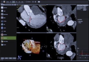
  7. Roy, et al. Novel Integrated 3D Multi-Detector Computed Tomography and Fluoroscopy Fusion for Left Atrial Appendage Occlusion Procedures. Catheter Cardiovasc Interv 2017;Mar 17, DOI:10.1002/ccd.26998
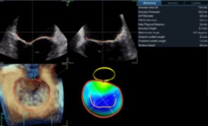
  8. Cvi42 mitral, distributed by GE for Circle Cardiovascular, is a leading application that offers a comprehensive toolset and workflow for the evaluation of the mitral valve. Accessible on AW 4,7 only. cvi mitral is sold separately. May not be available in all markets. Refer to your sales representative
  9. 15596_MVQ_Whitepaper_v7.pdf

JB05147XX