Sonographers are essential to delivering high-quality cardiac care, but their work comes with challenges. Repetitive manual steps, long exam times, and physical strain all take a toll. Echocardiography AI enables sonographers to focus on acquiring the best images while intelligent algorithms prepare measurements and draft reports. The result: high-quality echocardiograms with fewer clicks and less manual effort.
The hidden burden of clicks in echocardiography
For sonographers, performing a transthoracic echocardiogram (TTE) is more than acquiring images. It’s a taxing routine of positioning, freezing, tracing, measuring, labeling, and documenting. A single exam can involve nearly 200 clicks for image acquisition and measurements.1 Multiplied across a full day’s schedule and the daily clicks quickly reach the thousands.
This isn’t just a matter of inconvenience. Up to 90% of sonographers experience pain from work-related musculoskeletal disorders (WRMSDs)2, most often in the shoulders, neck, back, or wrists. These injuries are in addition to the cognitive fatigue of manual reporting and measurement, which can all contribute to burnout and attrition. For some, the cumulative toll results in extended sick leave or the decision to leave the profession altogether.
The effects ripple beyond an individual sonographer’s well-being. When skilled sonographers are sidelined, practices face reduced productivity amid existing turnover, recruitment challenges, and appointment backlogs. The Occupational Safety and Health Administration (OSHA), estimates that WRMSDs cost U.S. employers an about $120 billion annually in direct and indirect costs.3
The promise of AI in echocardiography
Artificial intelligence (AI) is reshaping ultrasound across specialties. From general radiology and obstetrics to cardiology, it is transforming how clinicians capture, interpret, and communicate diagnostic information. AI also emerging as a powerful enabler of efficiency and consistency across imaging workflows.
In echocardiography, these advancements are especially meaningful. Cardiac exams have unique attributes that make them complex and labor-intensive, so they demand particular precision and expertise. AI is increasingly capable of automatically detecting cardiac structures, calculating key measurements, and generating draft reports. These capabilities help ensure standardized results while reducing the manual workload that often slows clinicians down.
Supporting sonographers day-to-day with echocardiography AI
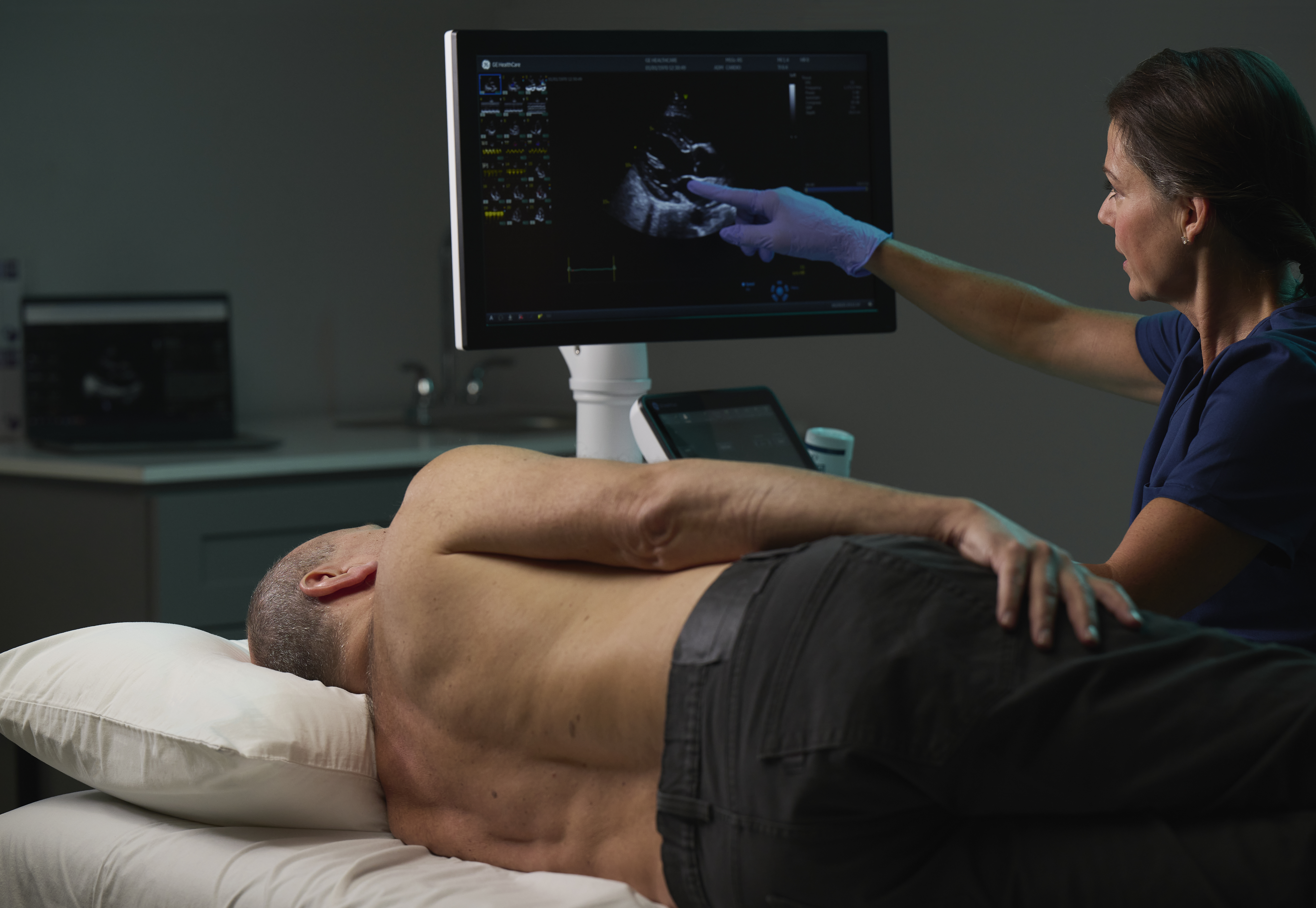
ViewPoint EchoPilot™, a cloud-based AI solution from GE HealthCare, brings this promise into daily echocardiography practice for cardiac sonographers. It reduces repetitive, manual steps throughout the exam and reporting process by:
- Automatically recognizing standard echocardiographic views and labeling them
- Detecting missing or incomplete views
- Calculating key cardiac measurements such as left ventricle ejection fraction, chamber dimensions and Doppler parameters
- Populating measurements and findings directly into a preliminary report
- Providing integrated access to ASE and EACVI reference ranges
- Generating a draft report so structured documentation is ready for review as soon as image acquisition is complete

ViewPoint EchoPilot means 65% fewer clicks, 50% faster scans
To evaluate the impact of ViewPoint EchoPilot, researchers compared two workflows for routine adult TTEs on the Vivid™ E95 ultrasound system. One protocol used a traditional manual process. The other integrated ViewPoint EchoPilot for automated measurement and reporting. Both workflows were tested on healthy male and female subjects aged 45–60, with no significant health conditions.
Results showed that a manual exam required about 200 clicks for measurements alone, while the ViewPoint EchoPilot workflow only needed 70, a 65% reduction.1 The automated workflow enabled clinicians to complete image acquisition and measurements in under 6 minutes, cutting scanning time in half.1

Explore the full infographic to see how ViewPoint EchoPilot™ helps sonographers reclaim time, reduce fatigue, and improve exam efficiency.
AI in echocardiography goes beyond efficiency metrics
For sonographers, the benefits of echocardiography AI are personal. By reducing the number of clicks and eliminating repetitive tracing tasks, AI lightens the physical workload that contributes to musculoskeletal injuries and day-to-day fatigue. This shift can help make the profession more sustainable, supporting sonographers in maintaining their health and career longevity.
At the same time, AI reduces the cognitive burden of manual reporting. With measurements prepared automatically, sonographers can devote more focus to capturing high-quality images and connecting with patients. Accuracy and consistency are built in, reducing rework and repeat exams. The impact on professional satisfaction could be considerable– a byproduct of feeling supported, and proud of delivering reliable results without repetitive manual effort.
Ready for the future of echocardiography
The combination of fewer clicks, faster exams, and consistent results sets a new benchmark for routine echocardiography. For sonographers, it may mean healthier, more rewarding workdays. For practice leaders, expanded patient access, stronger retention, and sustainable growth are feasible.
As demand for cardiac imaging grows, innovations like ViewPoint EchoPilot will be essential. Echocardiography AI helps ensure that sonographers can perform more exams without added strain, enabling clinicians and patients alike to benefit from a smarter, more sustainable workflow.
REFERENCES:
- 1.Based on a single internal, side-by-side comparison study performed by GE HealthCare. Three certified sonographers completed a routine adult transthoracic echocardiogram on the Vivid E95 system using two distinct workflows: a traditional manual and an automated workflow using ViewPoint EchoPilot. In the manual workflow, sonographers manually acquired images and performed measurements, followed by report generation in ViewPoint 6. No automation capabilities available on the scanner were utilized during this process to ensure a true baseline comparison. All final reports produced through the automated workflow still would require final clinical interpretation and validation by a qualified physician. Consistent number of images, measurements, and exam details across both workflows were performed. Live scanning was conducted on external scan subjects, including both male and female participants aged 45-60 years, with no significant health conditions. A total of 18 data sets were collected-9 per protocol-including both scan data and completed reports.
- 2.Work-related Musculoskeletal Disorders (WRMSD) Grand Challenge. American Registry for Diagnostic Medical Sonography 2025. Available at: https://www.ardms.org/wrmsd-grand-challenge/. Accessed October 15, 2025.
- 3.Prevention of Work-Related Musculoskeletal Disorders. Occupational Safety and Health Administration 2016. Available at: https://www.osha.gov/pls/oshaweb/owadisp.show_document?p_table=UNIFIED_AGENDA&p_id=4481. Accessed July 8, 2016.
ViewPoint, Vivid and EchoPilot are trademarks of GE HealthCare.
Product and features may not be available in all countries and regions. Full product technical specification is available upon request. Contact a GE HealthCare representative for more information.
©2025 GE HealthCare. GE is a trademark of General Electric Company used under trademark license.
JB35604XX